MRSIPrep: A Robust Preprocessing Pipeline for Whole-Brain MRSI Data
MRSIPrep is a preprocessing and derivative-generation pipeline for already quantified whole-brain MRSI maps, run as a BIDS App via Docker.






About
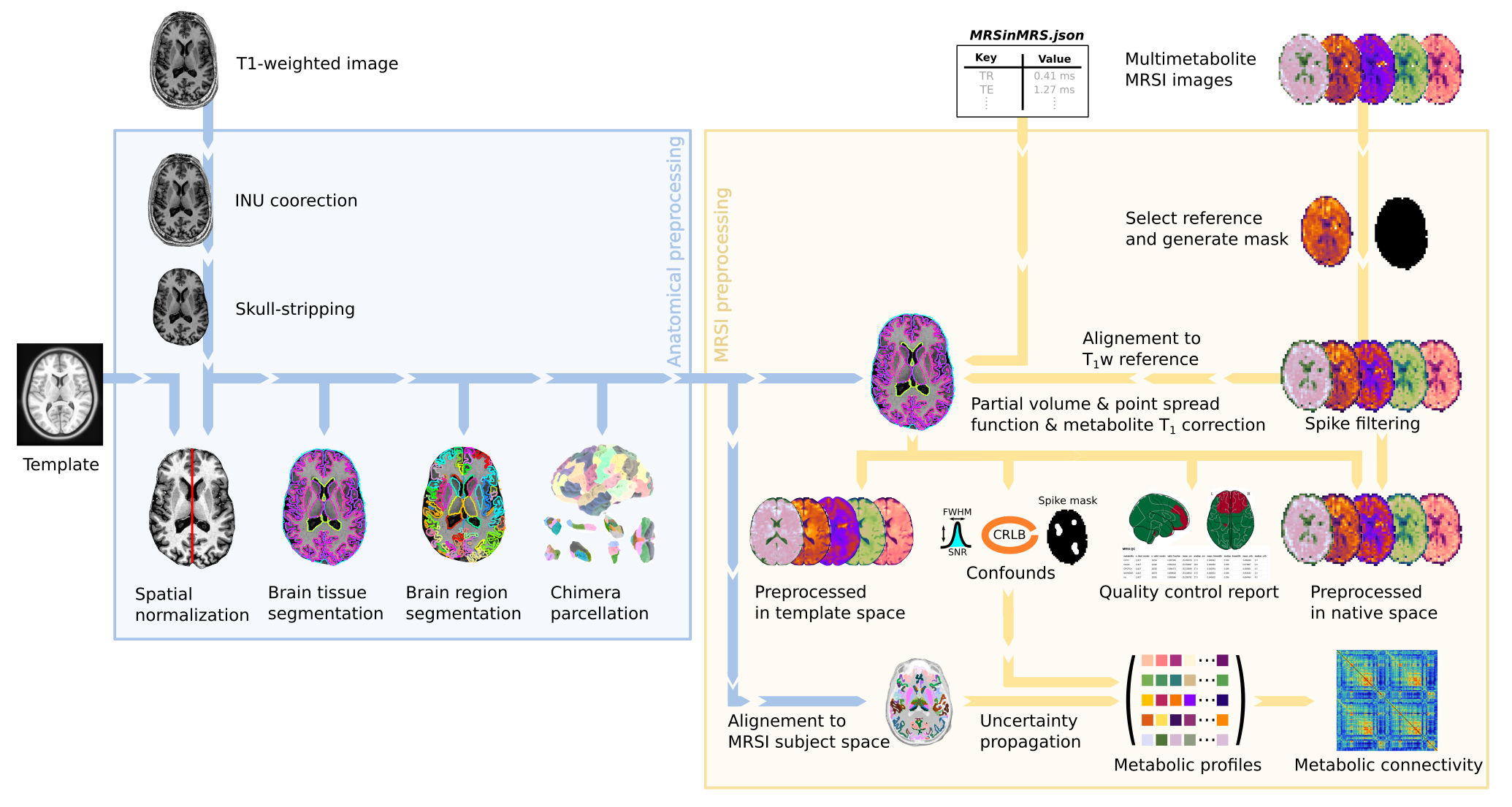
MRSIPrep is a magnetic resonance spectroscopic imaging (MRSI) data
preprocessing pipeline that is designed to provide an easily accessible,
robust interface requiring minimal user input, while providing easily
interpretable and comprehensive quality-control reporting. It performs basic
processing steps (registration to anatomical and template space, tissue
segmentation, partial-volume correction, atlas projection, etc.) on already
quantified whole-brain MRSI maps, providing outputs that can be easily
submitted to a variety of group-level analyses, including
voxel-based analysis, regional
statistics, and metabolic connectivity.
It is derived from the implementation in MRSI-Metabolic-Connectome and preserves the CHUV
academic non-commercial research license.
Every report includes the acquisition's MRSinMRS
minimum-reporting-standard sequence parameters (read from an optional
mrsinmrs.json at the BIDS root) and a citation section, so results are
easy to trace back to both the software and the acquisition protocol used.
Published-study parameter sets can be reproduced directly with
--config-preset <name> (see --list-presets); the report then credits
the source publication.
Test Dataset
A small, public, synthetic MRSI dataset — SynthMRSI-Project — is
available for anyone to download and run through MRSIPrep themselves,
without needing access to real MRSI acquisitions. It pairs real T1w
anatomical images (subsetted from two CC0 OpenNeuro datasets) with
model-synthesized MRSI signal and empirical CRLB/SNR/FWHM quality maps,
following MRSIPrep's own raw-MRSI-input convention
(derivatives/mrsi-orig/).
Published on Zenodo: 10.5281/zenodo.21477047 (CC0). See PUBLIC_DATASET.md in the repository for full download and usage instructions. This dataset is also the fixture for MRSIPrep's own automated end-to-end pipeline test.
What it uses
Nipype as the workflow engine: each subject/session is a cached, per-step Nipype workflow, so a rerun of an already-processed recording skips finished steps instead of recomputing them.
ANTs for MRSI↔T1w and T1w↔MNI registration.
FreeSurfer (
mri_synthseg,recon-all,mri_vol2vol) for brain extraction, cortical/subcortical parcellation, and surface reconstruction.FSL FAST for tissue-class probability segmentation.
PETPVC for partial-volume correction of MRSI maps.
Chimera for multi-atlas cortical/subcortical parcellation fusion.
TemplateFlow for the bundled MNI152 reference templates and atlases.
Pipelines
MRSIPrep runs in one of two modes, selected with --mode:
mni-norm(MNI normalization, default) — registers MRSI maps to a SynthSeg-extracted T1w image, generates SynthSeg+FAST tissue probability maps, applies PETPVC partial-volume correction (--no-pvcto disable), resamples to the requested output spaces, and parcellates with SynthSeg cortical/subcortical labels. No Chimera, norecon-all. This is the fast default path for anatomical coverage and CRLB reporting.parc-con(parcellation and connectivity) — adds a choice of Chimera multi-atlas or bundled MNI-atlas parcellation, plus optional perturbation-based connectivity matrices.
Both modes share the same MRSI filtering, quality-masking, and T1w/MNI
normalization machinery; parc-con mode is a superset of mni-norm outputs.
Design Principles
MRSIPrep was designed according to four main principles: reproducibility, modularity, transparency, and analysis agnosticism.
Reproducibility
The framework is distributed as open-source software and can be executed in containerized environments to minimize differences across computing platforms.
Modularity
Each processing stage is implemented as an independent module, allowing users to enable, disable, or replace specific steps according to their acquisition protocol and scientific question.
Transparency
MRSIPrep generates automated quality-control reports summarizing spatial registration, metabolite coverage, voxel-level quality metrics, tissue composition, and atlas projection.
Analysis Agnosticism
MRSIPrep does not impose a specific downstream analysis. Instead, it generates standardized derivatives that can be used for voxelwise analyses, regional analyses, metabolic connectomics, gradient mapping, or machine-learning workflows.
Workflow Architecture
Inputs
MRSIPrep starts from quantified metabolite maps and associated quality metrics. Typical inputs include metabolite concentration maps, Cramér-Rao lower bound maps, signal-to-noise ratio maps, linewidth maps, anatomical T1-weighted images, tissue probability maps, and optional atlas files.
Processing Steps
The core processing workflow includes:
MRSI-BIDS-compatible data import.
Voxelwise quality assessment.
Brain masking and coverage estimation.
Tissue fraction estimation.
CSF and tissue correction.
Spatial registration to anatomical and template spaces.
Atlas projection and regional summary extraction.
Generation of voxelwise, regional, and connectomics-ready derivatives.
Automated quality-control reporting.
Quality-Control Framework
MRSIPrep summarizes quality at the voxel, regional, and subject levels. Voxel inclusion can be based on metabolite-specific criteria such as linewidth, signal-to-noise ratio, Cramér-Rao lower bounds, tissue composition, and spatial coverage.
License
MRSIPrep is distributed under the CHUV academic non-commercial research license; see LICENSE for the full text.
Acknowledgments
Substantial implementation logic is cropped and refactored by Federico
Lucchetti and Edgar Céléreau from MRSI-Metabolic-Connectome. MRSIPrep
builds on the work of the ANTs, FreeSurfer, FSL, PETPVC, Chimera, and
TemplateFlow projects.
Publications
Code derived from this pipeline has been used in the following peer-reviewed publications:
Lucchetti, F., Céléreau, E., Steullet, P., Alemán-Gómez, Y., Hagmann, P., Klauser, A., & Klauser, P. (2025). Constructing the human brain metabolic connectome with MR spectroscopic imaging reveals cerebral biochemical organization. Nature Communications, 16. doi:10.1038/s41467-025-66124-w
Céléreau, E., Lucchetti, F., Alemán-Gómez, Y., Dwir, D., Cleusix, M., Ledoux, J.-B., Jenni, R., Conchon, C., Bach Cuadra, M., Schilliger, Z., Solida, A., Armando, M., Plessen, K. J., Hagmann, P., Conus, P., Klauser, A., & Klauser, P. (2026). High-resolution whole-brain magnetic resonance spectroscopic imaging in youth at risk for psychosis. Imaging Neuroscience, 4. doi:10.1162/imag.a.1276